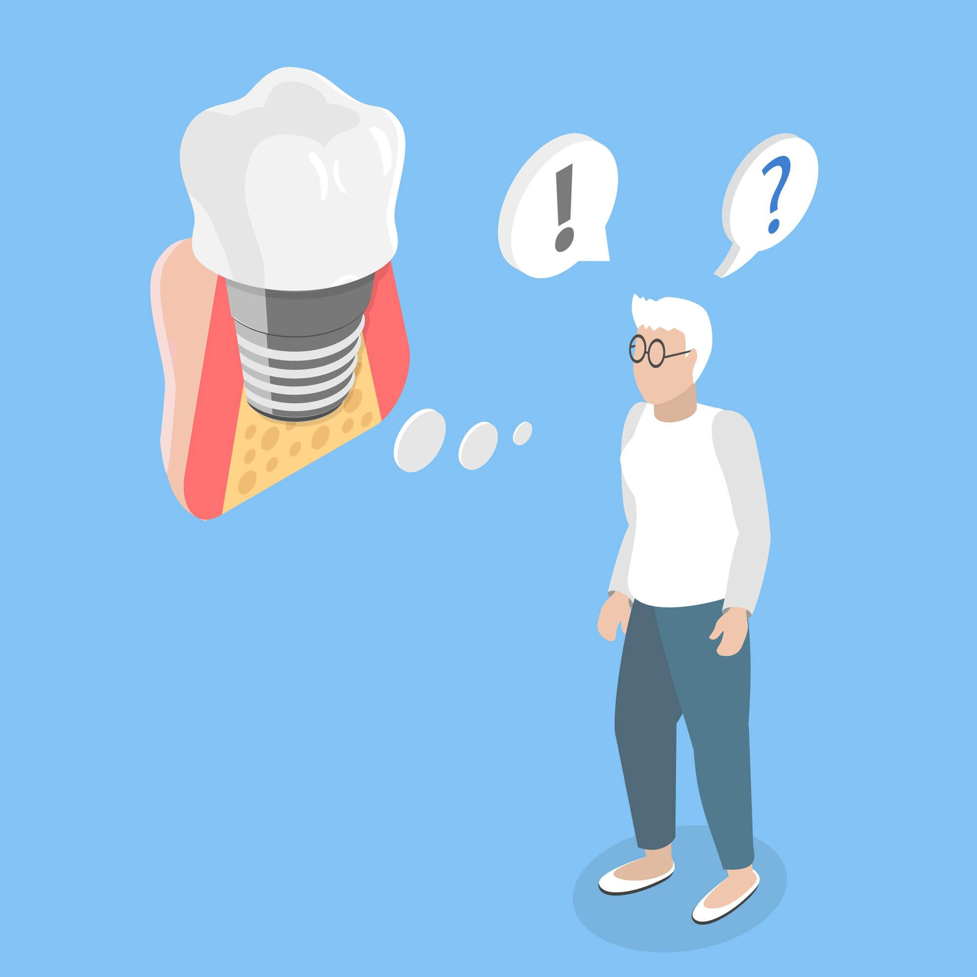
Dental implants can fail when the implant does not fuse properly with the jawbone, when infection damages the surrounding tissue, or when too much pressure affects the implant, abutment, or crown. Most modern implants have high long-term success rates, often reported between 90% and 98% over 10 years in suitable candidates, yet failure is still possible. The reason is rarely random. It usually relates to bone healing, oral hygiene, smoking, uncontrolled medical conditions, bite pressure, or technical issues in planning and placement.
Failure may happen early, often within the first 3 to 6 months, when osseointegration does not occur as expected. In these cases, the implant may become loose, painful, or show poor healing. Failure can also happen later, sometimes years after successful placement, when bone loss develops around the implant. This later pattern is frequently linked to peri-implantitis, which is an inflammatory condition caused by bacterial buildup around the implant, or by excessive biting forces and mechanical wear.
Common warning signs include persistent pain after the expected healing window, swelling, bleeding around the implant, gum recession, a loose implant or crown, bad taste, pus, or difficulty chewing. Some failed implants can be treated if the problem is limited to the crown, abutment, bite alignment, or early inflammation. If the implant itself is loose, removal is often necessary. After treatment of infection or bone loss, many patients can have another implant placed, sometimes after bone grafting and a healing period of 3 to 6 months.
Careful diagnosis matters. Dentists use clinical exams, X-rays, 3D CBCT scans, pocket depth measurements, and bite analysis to find the exact cause. The most effective way to reduce failure risk is good case selection, precise implant positioning, daily brushing and interdental cleaning, regular maintenance visits every 4 to 6 months for higher-risk patients, and strict control of smoking, diabetes, and teeth grinding.
Common Causes of Dental Implant Failure
Dental implant failure usually develops from biological problems, mechanical stress, or treatment-planning issues. Even though implants are designed to function for many years, they depend on healthy bone, stable gums, controlled bite forces, and consistent home care. When one or more of these conditions are missing, the likelihood of failure rises. In routine practice, the most frequently discussed causes are failed osseointegration, infection, peri-implantitis, overload from chewing or grinding, poor implant positioning, and insufficient bone support.
Osseointegration is the process in which the implant surface bonds with the jawbone. This stage often takes about 8 to 16 weeks in straightforward cases, though some patients need longer healing. If the bone does not integrate with the implant, the fixture may stay mobile or become painful. This problem can be linked to low bone density, overheating of bone during drilling, early loading, uncontrolled diabetes, heavy smoking, or micromovement greater than roughly 50 to 100 microns during healing. A stable implant needs a controlled surgical environment and a healing period that matches the patient’s bone quality.
Infection is one of the clearest causes of implant complications. Early infection can occur soon after surgery if bacteria enter the wound or if oral hygiene is poor during healing. A later form of infection, peri-implantitis, involves inflammation and bone loss around an implant that had once been stable. This condition is often compared with advanced gum disease around natural teeth. Bleeding on probing, pocket depths above 5 or 6 mm, radiographic bone loss, and suppuration can all point to peri-implantitis. In some long-term studies, peri-implantitis affects roughly 10% to 20% of implant patients, though the exact number depends on the patient population and the definition used.
Excessive force is another common cause. Dental implants do not have the periodontal ligament that natural teeth have, so they do not absorb pressure in quite the same way. If an implant crown is too high, if the bite is uneven, or if the patient clenches or grinds at night, stress can build up. That may lead to screw loosening, crown fracture, abutment problems, or gradual bone loss around the implant neck. Bruxism can increase occlusal load significantly, and without a night guard in selected cases, repeated strain can shorten implant survival.
Poor treatment planning can also set the stage for failure. An implant placed in a position with limited bone thickness, too close to adjacent teeth, too close to another implant, or in an angle that creates cleaning difficulty may function poorly over time. Inadequate prosthetic design matters as well. A bridge or crown that traps plaque, has excess cement beneath the gumline, or distributes force badly can increase inflammation and bone loss. Cement-related peri-implant disease is a well-known issue, which is why many clinicians now favor screw-retained restorations in selected cases.
Systemic and local healing factors often overlap with the causes above. Patients with uncontrolled diabetes, severe periodontal disease, dry mouth, immune compromise, head and neck radiation history, or long-term antiresorptive medication use may face more complex healing patterns. A dental implant does not fail for one reason in every case. It usually reflects a combination of tissue health, bacterial control, biomechanics, and planning accuracy.
Early vs Late Dental Implant Failure
Dental implant failure is often grouped into early failure and late failure because the timing gives useful clues about the cause. Early failure usually happens before the implant has fully integrated with the bone. In many cases, this means the first few weeks or months after surgery, often within 3 to 6 months. Late failure happens after the implant initially healed and functioned well, sometimes after 1 year, 5 years, or even longer. The difference is not only about timing. The biological processes, treatment approach, and prognosis can be quite different.
Early implant failure is commonly related to failed osseointegration. The implant may never develop a stable bond with the jawbone. This can happen if the surgical site was infected, if the implant lacked primary stability, if the bone quality was poor, or if excessive movement occurred during healing. Smoking more than 10 cigarettes a day, uncontrolled diabetes with elevated HbA1c levels, untreated periodontal infection, or immediate loading in an unsuitable case can all contribute. A patient may notice unusual pain, mobility, persistent swelling, or delayed healing. Some early failures are only detected during the restorative phase when the dentist tests the implant and finds that it is not stable enough to support a crown.
Late implant failure tends to develop after a period of apparent success. A patient may have used the implant comfortably for years before signs of trouble appear. The most common reasons are peri-implantitis, progressive bone loss, and chronic overload. Peri-implantitis begins with bacterial inflammation in the soft tissue and can progress into bone destruction around the implant threads. If this process is not managed, the implant may loosen. Late failure can also be mechanical. A loose abutment screw, repeated porcelain fracture, or poorly distributed chewing forces may not destroy the implant immediately, but they can create conditions that damage the surrounding tissue over time.
The symptoms can overlap, yet there are patterns. Early failure often looks like non-healing: tenderness that does not settle, the implant feeling unstable, or the area not progressing as expected by week 2, week 6, or month 3. Late failure may start more quietly with bleeding while brushing, mild gum swelling, a bad taste, deeper pockets, or slight bone loss seen on routine X-rays. Some patients do not feel pain until the condition is already advanced.
Management also differs. An early failed implant is often removed, the site is cleaned, and the bone is allowed to heal for several weeks or months. Re-implantation may be possible after reassessment of bone volume and risk factors. Late failure may sometimes be managed without removing the implant if the issue is limited to inflammation, excess cement, crown design, or bite overload. If bone loss is advanced and the implant is mobile, removal is usually recommended. Distinguishing early from late failure helps the dentist trace the cause and build a safer plan for replacement.
Signs and Symptoms of a Failed Dental Implant
A failed dental implant does not always cause dramatic symptoms in the beginning. Some cases start with subtle changes that are easy to miss, which is why regular follow-up matters. The most important sign is mobility of the implant itself. A healthy implant should feel rigid because it is fused to bone. If the implant body moves, that is a strong warning sign of failure. Patients sometimes confuse a loose crown or abutment with a loose implant, so a clinical exam is needed to identify which part is moving.
Pain is another key symptom, though the timing matters. Some soreness is expected after implant surgery for a few days, and mild tenderness can continue for 1 to 2 weeks. Persistent pain beyond the expected healing period, pain that returns after the area had settled, or discomfort during chewing months later can indicate a problem. Sharp pressure pain may point to overload or inflammation. Throbbing pain may suggest infection. A stable implant should not cause daily pain once healing is complete.
Swelling, redness, and bleeding around the implant are common soft tissue warning signs. Healthy gum tissue around an implant should look pink and firm, not puffy or shiny. If brushing causes regular bleeding, if there is spontaneous bleeding, or if the gums look inflamed for more than a few days, peri-implant mucositis or peri-implantitis may be present. Pocketing around implants can become deeper over time, and in a dental exam, probing depths above 5 mm with bleeding or pus are often treated seriously.
Pus or discharge is a more urgent symptom. A bad taste in the mouth, repeated fluid release when the gums are pressed, or a small pimple-like opening near the implant can signal active infection. This may be accompanied by a foul smell, tenderness, and progressive bone loss. If infection is allowed to continue, the bone supporting the implant can shrink enough to make the implant unstable.
Gum recession can also be a visible sign. If the gumline drops and more of the implant or crown margin becomes exposed, the area can trap plaque more easily and may reflect underlying bone loss. In the front of the mouth, patients often notice cosmetic changes before discomfort. In the back of the mouth, chewing difficulty may be the first clue.
Functional changes matter as much as visible ones. If biting feels uneven, if the implant crown hits first, if food constantly packs around it, or if the crown suddenly feels taller than before, a bite or hardware problem may be developing. Clicking or slight movement can suggest a loose screw rather than full implant failure, but this still needs prompt care because delayed repair can lead to fracture or tissue damage.
Many failed implants are discovered on routine X-rays before symptoms become severe. Marginal bone loss of about 1 to 1.5 mm in the first year can be within normal remodeling limits, followed by much smaller annual changes. Bone loss that is progressive, irregular, or paired with inflammation raises concern. Any implant that feels different from the surrounding teeth for more than a few days deserves assessment.
Risk Factors That Increase the Chance of Implant Failure
Dental implant failure becomes more likely when several risk factors overlap. One factor does not guarantee failure, yet the odds change when bone quality, general health, oral hygiene, smoking, and bite pressure all work against healing. Some risks are patient-related, some are disease-related, and some are linked to treatment design. Knowing these variables helps patients and clinicians make realistic decisions before surgery.
Smoking is one of the best-known risks. Nicotine reduces blood flow, affects soft tissue healing, and can interfere with bone formation around the implant surface. Heavy smokers often show higher rates of early healing problems and later peri-implant bone loss. Studies vary, though many report implant complications to be noticeably higher in smokers than in non-smokers. The risk tends to rise with the number of cigarettes per day and with long-term tobacco use. Vaping is not automatically safe in this context either, since nicotine and heat exposure may still affect healing.
Uncontrolled diabetes is another major factor. Patients with well-managed diabetes can often receive implants successfully, but poor glycemic control is associated with slower healing, infection risk, and reduced osseointegration quality. Many clinicians pay close attention to HbA1c levels before elective implant surgery. A patient with an HbA1c around 7% or below may heal more predictably than someone with substantially higher levels, though the full medical picture always matters.
History of periodontal disease increases long-term risk. If a patient has lost teeth due to chronic gum disease, the bacteria and inflammatory patterns that damaged natural teeth can also threaten implants. This does not mean implants should be avoided, but maintenance usually needs to be stricter. Professional cleaning intervals may need to be every 3 to 4 months instead of every 6 months, and home care must be consistent.
Insufficient bone volume or low bone density can reduce primary stability. The upper back jaw often has softer bone than the lower front jaw, which can make integration more challenging. Bone grafting, sinus lift procedures, or longer healing times may be recommended when the site is not ideal. Skipping these steps in a borderline case can increase the chance of failure.
Bruxism, clenching, and heavy bite forces place implants under repeated stress. Natural teeth have a small cushioning ligament. Implants do not. A person who grinds at night may generate forces strong enough to loosen screws, chip crowns, or contribute to marginal bone loss. A custom night guard is commonly recommended in these patients.
Certain medications and medical treatments also deserve attention. Bisphosphonates and other antiresorptive drugs, high-dose steroids, some cancer therapies, and past radiation therapy to the jaw can affect bone turnover or healing response. Dry mouth from medications can worsen plaque accumulation. Immune disorders may also complicate healing.
Poor oral hygiene remains a simple but powerful risk. Plaque retention around implants can trigger mucositis within weeks and peri-implantitis over time. Inadequate cleaning is more likely when crown contours are bulky, access is tight, or the patient has limited dexterity. Implant design, prosthetic design, and maintenance habits all influence whether bacteria can be controlled day after day.
Can a Failed Dental Implant Be Saved?
Some failed dental implants can be saved, though the answer depends on what has actually failed. Patients often use the phrase “failed implant” to describe any problem around an implant, but the treatment path changes a lot depending on whether the issue is a loose crown, a loose abutment screw, gum inflammation, bone loss, or a truly loose implant fixture. If the implant body is still firmly integrated in bone, there may be a good chance of preserving it. If the implant itself is mobile, saving it is much less likely.
One of the more treatable situations is a prosthetic problem rather than implant failure. A crown can loosen, a screw can back out, or the bite can become imbalanced. In these cases, the dentist may remove the crown, inspect the abutment, tighten or replace the screw, adjust the bite, and check the surrounding tissues. This can often be done in a single visit of 30 to 60 minutes, though replacement parts or a new crown may extend the process. When the supporting bone remains healthy, the prognosis is often favorable.
Early inflammatory problems can also be reversible. Peri-implant mucositis affects the soft tissue without significant bone loss. If caught early, it may respond to professional cleaning, improved home care, antimicrobial rinses in selected cases, and correction of plaque-retentive contours. Bleeding and swelling may settle within a few weeks if the source is controlled. Once bone loss develops, the condition is no longer a simple soft tissue issue.
Peri-implantitis is harder to save but not always hopeless. Management may include mechanical debridement, removal of excess cement, antiseptic treatment, laser or air-abrasive decontamination, systemic or local antibiotics when indicated, and surgical access to clean the implant surface. In some cases, regenerative procedures using bone graft and membranes are considered, though outcomes vary based on defect shape and implant surface condition. Implants with mild to moderate bone loss and no mobility have a better chance of remaining in service than implants with advanced circumferential loss.
An implant that is mobile usually cannot be saved. Mobility means osseointegration has failed or has been lost to the point that the implant no longer has stable support. Leaving a mobile implant in place can worsen bone destruction and prolong infection. Removal is normally the safer option. The site may then be grafted and allowed to heal for 8 to 16 weeks or longer, depending on bone damage, before deciding whether to place a new implant.
Timing makes a real difference. A patient who reports bleeding, bad taste, swelling, or pressure discomfort early may avoid a more invasive procedure later. When our readers ask whether a failed implant can be rescued, the most honest answer is that some can, some cannot, and the deciding factors are mobility, amount of bone loss, infection control, and how fast the issue is addressed.
Treatment Options for a Failed Dental Implant
Treatment for a failed dental implant depends on whether the problem is mechanical, inflammatory, or structural. There is no single protocol for every case. The dentist has to identify which component is involved, how much supporting bone remains, whether infection is active, and whether the implant is still stable. This is why two patients with similar symptoms may receive very different plans.
Minor prosthetic issues are often the simplest to treat. If the crown is loose but the implant is stable, the dentist may remove the restoration, inspect the screw channel, tighten or replace the abutment screw, and adjust the bite. If the crown is cracked, chipped, or poorly shaped, a new crown may be made. Costs vary widely by country and clinic, though replacing an implant crown may range from a few hundred to over $1,500, while a simple screw-retightening visit is much lower. These problems are inconvenient, yet they do not always mean implant failure.
When soft tissue inflammation is present without major bone loss, treatment may focus on peri-implant mucositis. Professional cleaning around the implant, polishing with implant-safe instruments, oral hygiene instruction, and short-term antimicrobial support may be used. Patients are often reviewed after 2 to 6 weeks to check whether bleeding and swelling have decreased. If plaque control improves and the restoration allows proper cleaning, the tissues may stabilize.
Peri-implantitis usually requires a more involved approach. Non-surgical treatment may help reduce inflammation, though deeper defects are often difficult to clean thoroughly without surgery. Surgical options can include flap access, removal of granulation tissue, implant surface decontamination, reshaping of the bone in some cases, or regenerative procedures with graft material and membranes. Follow-up is crucial because recurrence can occur if the patient has ongoing plaque issues, smoking habits, or uncontrolled diabetes.
If the implant is mobile, fractured, severely malpositioned, or associated with extensive bone loss, removal is often necessary. Implant removal can be straightforward in some cases and more complex in others, depending on the degree of integration and the amount of surrounding bone. The site may be curetted, disinfected, and grafted. Bone graft materials may include allograft, xenograft, synthetic substitutes, or combinations. Healing commonly takes about 3 to 6 months before reassessment, though larger defects can require longer.
Replacement after removal is often possible. Some patients receive a new implant in the same site after graft healing. Others may need a wider or longer implant, a different position, or a staged plan with sinus augmentation or ridge rebuilding. If a repeat implant is not ideal, alternatives include a conventional bridge, a resin-bonded bridge in selected front-tooth cases, or a removable partial denture. The best option depends on the amount of remaining bone, esthetic goals, medical status, budget, and how much treatment time the patient can accept.
Many patients worry that one failed implant means all future implants will fail. That is not automatically true. If the cause is identified and corrected, re-treatment can succeed. The key is to rebuild the plan around the reason the first implant failed rather than simply repeating the same approach.
How Dentists Diagnose Dental Implant Failure
Diagnosing dental implant failure involves more than checking whether an implant feels loose. Dentists look at symptoms, gum condition, radiographic bone levels, the stability of each component, and the patient’s medical and bite history. A proper diagnosis separates true implant failure from a loose crown, a screw problem, temporary gum irritation, or pain coming from a nearby tooth or sinus. This matters because treatment for each condition is different.
The process usually starts with a clinical examination. The dentist asks when the implant was placed, when symptoms began, whether the problem is constant or intermittent, and whether there is pain during chewing, brushing, or at rest. Bleeding, swelling, a bad taste, pus, or a feeling that the implant has become taller or unstable can offer important clues. Medical updates are also relevant. Changes in diabetes control, smoking habits, medications, or recent illness may help explain tissue breakdown.
Visual and tactile assessment comes next. The dentist checks the color and shape of the gum tissue, looks for recession, and tests whether plaque is collecting around the restoration. Gentle probing may be used to measure pocket depths and bleeding points. Around healthy implants, tissues should remain stable with minimal inflammation. Deeper pockets, suppuration, and tenderness raise suspicion for peri-implant disease. Mobility testing is critical. If only the crown or abutment moves, the implant body may still be stable. If the fixture itself moves, the prognosis is much worse.
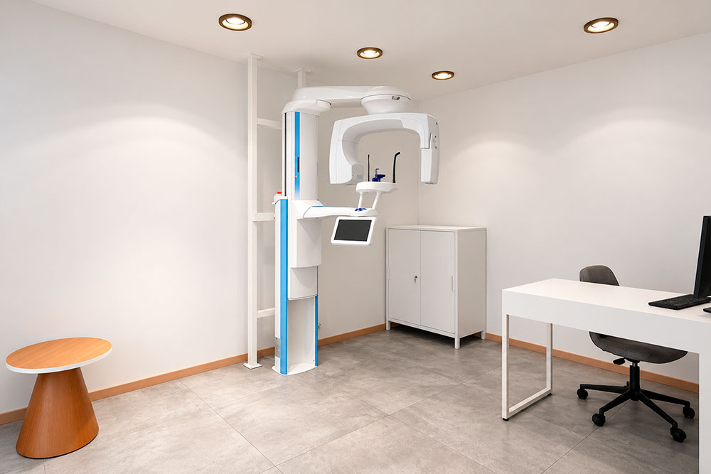
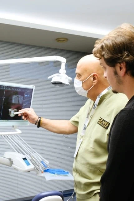
Radiographs are central to diagnosis. Standard periapical X-rays help measure crestal bone levels and compare the implant to earlier records. If bone loss appears progressive or if anatomy is unclear, a CBCT scan may be recommended. CBCT provides a 3D view that can reveal crater-like defects, buccal or lingual bone loss, proximity to the sinus or nerve, and implant angulation issues. This is particularly useful in complex re-treatment cases. A small amount of bone remodeling after placement can be normal, yet progressive vertical or circumferential loss is not.
Occlusal analysis is often overlooked by patients but important clinically. The dentist may use articulating paper, shimstock, or digital tools to see whether the implant crown is taking excessive force. Night grinding signs on other teeth can support the diagnosis of overload. If screw loosening has happened more than once, force distribution becomes a major concern.
In some cases, the restoration is removed to inspect the abutment, screw head, and implant connection. This can reveal hidden problems such as screw fracture, stripped threads, excess cement, or poor fit. If infection is suspected, the dentist may document probing depths and radiographic changes over time rather than relying on one single visit. Diagnosis is often built from several findings together: symptoms, soft tissue inflammation, bone loss pattern, and mechanical stability. That full picture is what guides whether the implant can be maintained, repaired, or removed.
How to Prevent Dental Implant Failure
Preventing dental implant failure starts before the implant is ever placed. A successful result depends on planning, surgical technique, healing conditions, prosthetic design, and the patient’s daily care routine. Long-term implant survival is not only about the day of surgery. It is built over months and years of maintenance.
Careful assessment is the first protective step. Dentists review bone quantity, gum health, bite forces, medical history, smoking status, and expectations before planning treatment. In many clinics, CBCT imaging is used to measure available bone in millimeters and avoid nerves or sinus spaces. If the site has only 4 to 5 mm of width where more is needed, grafting may be advised rather than forcing placement into a compromised position. Treating gum disease before implant surgery is also important because active periodontal infection can increase bacterial risk around the new implant.
Patients can reduce failure risk by controlling modifiable health factors. Smoking cessation is one of the most valuable changes. Even cutting down helps less than complete stopping, though any reduction may improve tissue response. Diabetes management matters too. A stable HbA1c level gives the body a better chance to heal predictably. If a patient grinds or clenches, a custom occlusal guard may protect the implant from repeated overload, often worn nightly.
Home care needs to be consistent and technique-specific. Implants do not get cavities, but the surrounding gums and bone can still become diseased. Brushing twice a day for about 2 minutes, cleaning between teeth daily with floss, interdental brushes, or water flossers where appropriate, and keeping plaque away from the implant margin are core habits. Patients with bridges or larger implant restorations may need special threaders or angled brushes to reach under the prosthesis.
Professional maintenance supports what home care cannot fully achieve. Many implant patients do well with hygiene visits every 6 months, though high-risk patients often benefit from 3- to 4-month intervals. During these visits, the dentist or hygienist checks bleeding, pocket depths, bite contacts, screw stability, and radiographic bone levels when indicated. Catching mucositis early can prevent more destructive peri-implantitis.
Prosthetic design also affects prevention. Crowns should allow access for cleaning and should not trap food or excess cement under the gums. The bite should be balanced so the implant does not take more force than intended. In full-arch cases, maintenance can be more involved, and patients need clear instructions about recall schedules and hygiene tools.
Prevention is practical rather than mysterious. Good diagnosis, careful placement, healthy lifestyle choices, daily plaque control, and regular review visits give implants the best chance to last 10, 15, or 20 years or more. When one weak link is ignored, the risk climbs.
When to Call a Dentist About a Dental Implant
It is normal to have some discomfort after implant surgery, which can make patients unsure about when to call the dentist. Mild swelling for 2 to 3 days, tenderness for about a week, and gradual improvement are expected in many cases. What matters is whether symptoms are following a healing pattern or moving in the wrong direction. If pain, swelling, or bleeding is getting worse instead of better, the office should be contacted.
Call a dentist promptly if the implant feels loose. A stable implant should not move. Patients sometimes notice movement while chewing, flossing, or touching the area with the tongue. Even if the looseness turns out to be only the crown or screw, it needs assessment because repeated movement can damage the connection or surrounding tissue. A same-week appointment is usually reasonable, and some clinics prefer to see this within 24 to 48 hours.
Pain that continues beyond the expected healing period deserves attention. If discomfort is still significant after 7 to 14 days, if pain increases after initially improving, or if chewing becomes difficult weeks or months later, the cause should be checked. New pain around an old implant is not something to ignore. It may reflect overload, a crack in the restoration, infection, or bone loss.
Bleeding, swelling, and pus are strong reasons to call. Occasional slight bleeding from vigorous brushing may not always mean failure, yet repeated bleeding around one implant, swollen gums, a bad taste, or visible discharge are signs that peri-implant inflammation may be active. Fever, facial swelling, or difficulty opening the mouth after surgery can indicate a more urgent infection and should be reported quickly.
Changes in appearance also matter. If the gum around the implant is receding, if metal or dark structure is becoming visible, or if the crown suddenly looks longer, bone and gum support may be changing. In front teeth, even a 1 to 2 mm shift can be noticeable and can become more difficult to correct if left untreated.
Functional changes should not be dismissed. Food trapping around the implant, a sensation that the crown hits first when biting, clicking, repeated screw loosening, or a crown that feels slightly lifted are all reasons to seek review. These issues may sound small, but they can progress into fracture, inflammation, or bone stress over time.
If a patient has risk factors such as smoking, prior gum disease, diabetes, or heavy grinding, a lower threshold for calling is sensible. Many implant complications are easier to manage when caught early. If the area feels different, looks inflamed, or functions differently for more than a few days, scheduling an exam is usually the safer choice.
FAQs About Dental Implant Failure
Patients often have practical questions about implant failure because the term covers many different problems. Some are minor repairs, while others require removal and replacement. Clear answers can help people respond quickly and avoid delay.
Can a dental implant fail years later? Yes. An implant can function well for many years and still develop trouble later. Late failure is often linked to peri-implantitis, chronic overload, or gradual bone loss. A 5-year-old or 10-year-old implant is not immune to infection if plaque control declines or if the bite becomes unfavorable.
Does pain always mean implant failure? No. Pain can come from gum irritation, a loose crown, a nearby tooth, clenching, or food impaction. A well-healed implant should not cause ongoing pain, though. Persistent or increasing discomfort needs evaluation.
If the crown is loose, has the implant failed? Not necessarily. A loose crown or abutment screw is a mechanical complication, not always a failed implant. If the implant fixture in the bone remains stable, the problem may be repairable without removing the implant.
What does a failed implant feel like? Patients may describe pressure pain, throbbing, tenderness during chewing, swelling, a bad taste, or a feeling that the implant moves. Some feel no pain and only notice bleeding or recession. Clinical examination is needed because symptoms overlap.
How common is dental implant failure? Exact rates vary by study and patient group, yet many modern implants show survival rates around 90% to 98% over 10 years in suitable conditions. Failure risk is higher in smokers, patients with poorly controlled diabetes, and those with a history of severe gum disease.
Can a failed implant be replaced? Often yes. After removal, the area may need bone grafting and a healing period of about 2 to 6 months before a new implant is placed. Some sites are ready sooner, while larger defects take longer.
How do dentists know whether the implant or just the screw is loose? The dentist checks each component clinically and may remove the crown to inspect the connection. X-rays help assess the surrounding bone. A loose screw can mimic implant mobility to a patient.
Can antibiotics fix a failed implant? Antibiotics alone rarely solve true implant failure. They may help control infection in selected cases, though they do not correct mobility, poor implant position, excess cement, or major bone loss.
Is bone loss around an implant always a sign of failure? No. Small early remodeling changes can be normal. Progressive bone loss with bleeding, pocketing, or pus is more concerning. Serial X-rays are useful because one image does not tell the whole story.
Can smoking really make that much difference? Yes. Smoking affects blood supply, wound healing, and tissue stability. In implant dentistry, it is one of the strongest modifiable risk factors.
What if the implant is fine but the gum is inflamed? That may be peri-implant mucositis, which can often be reversed with professional cleaning and better plaque control if caught early.
Is implant removal painful? Removal is usually done with local anesthesia, and many patients tolerate it well. Recovery depends on how integrated the implant is and whether grafting is done at the same visit.
Can grinding teeth damage implants? Yes. Bruxism can increase force on the crown, screw, abutment, and surrounding bone. A night guard is commonly advised when signs of grinding are present.
How often should implants be checked? Many patients are reviewed every 6 months, though higher-risk patients may need 3- to 4-month maintenance visits with periodic radiographs.
Should I wait if the implant only bleeds a little? Repeated bleeding around an implant is worth checking. Early inflammation is easier to manage than advanced peri-implantitis.