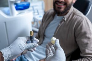
Most modern dental implants do not prevent you from having an MRI and are generally considered safe during scanning. In routine clinical practice, the majority of implants are made from titanium or titanium alloys, which are non-ferromagnetic or only weakly magnetic. That means they are not expected to move, twist, or become dangerous inside an MRI scanner that commonly operates at 1.5 Tesla or 3 Tesla. Many people with one implant, several implants, implant-supported crowns, or full-arch restorations undergo MRI without any complication.
The main issue is usually not safety but image quality. Metal in the mouth can sometimes create artifacts, which are areas of distortion or signal loss on MRI images. These artifacts are more likely to matter when the scan is focused on nearby anatomy such as the jaw, face, oral cavity, sinuses, temporomandibular joint, upper neck, or brainstem region. If the MRI is for a knee, spine, pelvis, or lower abdomen, dental implants rarely affect the diagnostic value of the exam.
It is still important to tell the MRI team about any implants, crowns, bridges, fixed retainers, or removable dental devices before the scan. The technician or radiologist may ask what type of material was used, when it was placed, and where it is located. If records are available, that information can help them plan the best scan settings. In many cases, the MRI can proceed as scheduled with no change. If artifacts are expected, the radiology team may adjust the sequence, angle, slice thickness, or positioning to reduce distortion.
For most patients, the practical answer is simple: dental implants are usually MRI-safe, but they can sometimes affect scans of the head and neck. A quick discussion with the imaging center and, if needed, your dentist or oral surgeon helps confirm the implant material and avoids surprises on the day of the exam.
Are Dental Implants Safe During an MRI?
Dental implants are usually safe during an MRI. Most implants placed over the last few decades are made from commercially pure titanium or titanium alloy, materials chosen for biocompatibility, corrosion resistance, and long-term stability in bone. Titanium is not strongly attracted to the magnetic field used in MRI, which is why it is widely accepted for use in orthopedic screws, joint components, craniofacial plates, and dental implant systems. In practical terms, this means a well-integrated implant in the jaw is not expected to shift or become dislodged during a standard scan.
MRI scanners in hospitals and imaging centers commonly operate at 1.5 Tesla or 3 Tesla. Safety concerns with metal generally relate to three areas: magnetic pull, heating, and image artifact. With modern dental implants, magnetic pull is typically negligible because titanium is non-ferromagnetic. Heating risk is also considered low. MRI can cause certain metallic objects to warm slightly due to radiofrequency energy, but small dental implants fixed inside bone do not usually reach clinically harmful temperatures. The risk profile is very different from loose metallic objects or devices with long conductive leads.
Clinical teams still screen every patient carefully because “metal in the body” is a broad category. Pacemakers, older aneurysm clips, neurostimulators, cochlear implants, insulin pumps, and some retained metal fragments raise different safety questions than dental implants. If a patient says, “I have metal in my mouth,” the MRI staff may ask whether it is an implant, crown, bridge, denture, orthodontic retainer, or surgical hardware. That distinction matters. A fixed titanium implant in the jaw is handled very differently from an unknown removable appliance.
Timing can matter in a few cases. A newly placed implant is mechanically stable when inserted and then gradually bonds to bone through osseointegration over roughly 6 to 12 weeks in many patients, though complete integration may continue for several months. Even so, MRI safety is generally tied more to the material than to the age of the implant. If placement was very recent and the patient has pain, swelling, or uncertainty about the device type, the imaging center may want supporting records before the scan.
Patients often worry because MRI sounds intense. The machine is loud, the environment is enclosed, and the magnetic field is powerful. Those facts can make any metal implant seem risky. From a dental and radiology standpoint, modern implants are among the more straightforward metallic items encountered in MRI screening. The key step is simple disclosure. Tell the MRI team what you have, where it is, and when it was placed. If you know the brand or material, bring that information. In most cases, the exam moves forward without a safety problem.
Can Dental Implants Interfere With MRI Results?
Dental implants can interfere with MRI results in some situations, but the effect depends heavily on what body part is being imaged and how close the implant is to the area of interest. MRI creates images by detecting signals from hydrogen atoms in tissues, and metal can disturb the local magnetic field. When that happens, the scan may show distortion, dark voids, signal loss, or warped anatomy near the metal. This is called artifact. The closer the implant is to the structure being studied, the more likely the artifact will matter clinically.
If the MRI is focused on the knee, lumbar spine, hip, ankle, or abdomen, dental implants almost never affect the final images in a meaningful way. The distance between the mouth and those regions is simply too great for oral metal to degrade image quality. If the MRI is aimed at the brain, pituitary area, sinuses, salivary glands, temporomandibular joints, oral cavity, tongue base, jawbone, or cervical spine, implants can become more relevant. In those cases, the metal may obscure a small portion of the field, and the radiologist may need to interpret around it or request modified sequences.
The extent of interference depends on several technical factors. Larger restorations tend to create more artifact than smaller ones. A single implant with a ceramic crown may have limited effect, while multiple implants, implant-supported bars, magnetic overdenture attachments, or dense cobalt-chromium components may generate more distortion. MRI field strength matters too. A 3 Tesla scanner can produce more susceptibility artifact than a 1.5 Tesla scanner in some scenarios. Pulse sequence selection also matters. Gradient-echo sequences are often more sensitive to metal than spin-echo-based techniques.
Radiology teams have ways to reduce interference. They may change the imaging plane, increase bandwidth, shorten echo time, switch sequences, or use dedicated metal artifact reduction techniques if available. Those adjustments can preserve diagnostic quality in many cases, though they may increase scan time by several minutes. A typical head MRI might last 20 to 35 minutes, while a more tailored protocol could extend to 40 or 45 minutes depending on the clinical question.
Interference is not the same as danger. Many patients hear “metal artifact” and assume the implant is unsafe. In reality, the concern is often whether the image remains clear enough for diagnosis. If the MRI is being ordered to evaluate a facial mass, jaw lesion, or inflammatory change near the mouth, even a small artifact can matter. If the purpose is to assess multiple sclerosis plaques, stroke, or inner ear structures, the effect may range from irrelevant to mildly limiting depending on implant position. This is why precise communication helps: when the radiologist knows exactly what dental hardware is present, the scan can be planned with fewer surprises.
What Types of Dental Implants Are MRI Compatible?
The dental implants most commonly considered MRI compatible are titanium and titanium-alloy endosteal implants. These are the standard screw-shaped implants placed in the jawbone to support crowns, bridges, or full-arch prostheses. Titanium dominates the global implant market because it combines strength, low weight, corrosion resistance, and excellent bone integration. In practical imaging terms, it is generally accepted as compatible with MRI conditions used in routine medical care, though it may still create localized artifact.
Zirconia implants are also relevant. Zirconia is a ceramic material rather than a metal, and it is often promoted for patients who prefer metal-free restorations or who prioritize esthetics in the front tooth region. From an MRI standpoint, zirconia tends to be highly favorable because it is non-metallic and less likely to create the same kind of magnetic susceptibility artifact seen with some metals. Zirconia implants are less common than titanium in many markets, yet they are a legitimate option. Their use may be limited by case selection, implant design, prosthetic flexibility, and clinician preference rather than MRI concerns.
Compatibility becomes less straightforward when the restoration includes more than the implant body. Some implant-supported crowns, abutments, bars, or attachments contain alloys such as cobalt-chromium, stainless steel components, or magnets used in certain overdenture systems. Those parts may remain safe but can create more artifact than titanium alone. Patients may say they “have implants,” when the imaging-relevant detail is actually the abutment or superstructure material. A fixed full-arch bridge on four or six implants can include a milled bar or framework made from materials that influence MRI appearance differently than the implants embedded in bone.
Older systems deserve extra attention. Dental implants placed 20 to 35 years ago may still be titanium, but records are sometimes incomplete and component materials may be less clear. Imported systems, custom prosthetic parts, and discontinued product lines can complicate identification. If the implant was placed in the last 10 to 15 years by a mainstream implant provider, documentation is often available and material identification is easier.
For patients preparing for MRI, the most useful compatibility information includes the implant brand if known, date of placement, number of implants, and whether there are any magnetic denture attachments or removable metal parts. The MRI team may not need every prosthetic detail for a scan of the knee or shoulder, but they may need it for a study of the face, jaw, or neck. In most routine cases, titanium and zirconia implant systems are handled without difficulty, with the main distinction being that zirconia tends to create less image disturbance while titanium remains the more common and clinically established choice.
Do Titanium Dental Implants Affect MRI Scans?
Titanium dental implants can affect MRI scans, but the effect is usually limited to image distortion near the implant rather than a safety problem. Titanium is the standard material for most dental implants because it is strong, lightweight, corrosion-resistant, and well tolerated by bone and soft tissue. In MRI, titanium is considered non-ferromagnetic or only weakly paramagnetic, which means it is not strongly pulled by the magnet the way ferromagnetic metals can be. That is why patients with titanium implants are commonly scanned in everyday medical practice.
The influence of titanium on the scan depends on what area is being imaged. If the MRI is for the lower back, pelvis, knee, or shoulder, titanium implants in the mouth will usually have no meaningful impact. If the MRI is directed at the face, oral cavity, paranasal sinuses, skull base, upper cervical spine, or nearby soft tissues, titanium can create susceptibility artifact. This often appears as a dark area, signal void, geometric distortion, or blurring of fine detail around the oral region. The degree of artifact can range from small and irrelevant to large enough to obscure the exact tissue a radiologist needs to evaluate.
Field strength plays a role. At 3 Tesla, artifact from titanium may appear somewhat more pronounced than at 1.5 Tesla, though modern sequence optimization can help. Scanner parameters such as bandwidth, voxel size, echo time, and pulse sequence selection all influence how much distortion is seen. Spin-echo and turbo spin-echo techniques often manage metal better than gradient-echo sequences. If the indication is highly specific, such as evaluating a lesion in the floor of the mouth or a mandibular nerve issue, the radiology team may tailor the protocol accordingly.
It also helps to separate the implant fixture from the restoration attached to it. A titanium implant buried in bone may produce modest artifact, while the abutment, screw, framework, or crown material can add to the effect. Porcelain-fused-to-metal restorations, cobalt-chromium structures, and dense metallic bars may alter images more than a single titanium fixture alone. Patients often assume the implant itself is the whole story, but the prosthetic assembly matters too.
From a practical standpoint, titanium implants affect MRI mainly when the area of interest is close to the mouth or jaw. They do not usually stop the scan from happening. If the exam is likely to be influenced, the MRI team may adjust the protocol, and in certain cases the referring doctor may choose another imaging method such as CT or cone beam CT for bone-focused questions. Titanium remains one of the most MRI-friendly materials used in implant dentistry, even though it is not completely invisible on scans.
Can Metal Dental Implants Cause MRI Artifacts?
Yes, metal dental implants can cause MRI artifacts, and this is one of the most common questions radiologists face when imaging the head and neck. An artifact is any image feature that does not represent normal anatomy or true pathology. With dental metals, the issue usually comes from distortion of the magnetic field around the object. That distortion changes how nearby tissue signals are recorded, producing dark bands, bright areas, geometric warping, or complete signal dropout.
The size and severity of artifact vary widely. A single small implant may create a limited area of disturbance measuring only a few millimeters to a couple of centimeters, depending on the scanner settings. A cluster of implants, a full-arch bar, metallic overdenture attachments, or dense alloy-based restorations may create a much larger region of degradation. The material matters a great deal. Titanium often causes less artifact than cobalt-chromium or some stainless-steel-containing components. Non-metal alternatives such as zirconia usually generate much less interference.
Where the artifact matters is just as important as how large it is. A mandibular implant may not affect imaging of the frontal lobes or upper chest, but it can interfere with assessment of the tongue, floor of the mouth, lower face, mandible, maxilla, and nearby lymph nodes. If the scan is looking for inflammation, soft tissue tumors, nerve involvement, osteomyelitis, or postoperative recurrence in those regions, metal artifact can reduce confidence in interpretation. A radiologist may still be able to answer the clinical question, though sometimes an alternate modality is preferred.
Artifact can often be reduced, though not always eliminated. Practical techniques include using fast spin echo sequences, increasing receiver bandwidth, decreasing voxel size, reducing echo time, adjusting the frequency-encoding direction, and applying metal artifact reduction software when available. These changes may improve image quality but can come with trade-offs such as lower signal-to-noise ratio or longer acquisition times. A protocol that would normally take 25 minutes may stretch to 35 or 40 minutes when optimization is needed.
Patients should know that artifact is common and usually manageable. It does not automatically mean the MRI failed. In many cases, the radiologist can still evaluate most of the anatomy and answer the referring doctor’s question. The significance depends on whether the area hidden by artifact overlaps with the region of concern. Clear communication before the scan gives the imaging team a better chance to plan around known dental hardware and obtain the most useful images possible.
Should You Tell the MRI Technician About Dental Implants?
Yes, you should tell the MRI technician about dental implants every time you attend an MRI appointment, even if you believe they are standard titanium implants and even if you have had MRI before. This is part of routine MRI safety screening. The technician, technologist, or radiographer needs an accurate list of implanted and removable items so the team can verify safety and anticipate image quality issues. Dental implants are common, and reporting them does not usually delay care, but not reporting them can create avoidable uncertainty once you are already in the scanner area.
The information does not need to be complicated. If you know the implant was placed in the upper jaw or lower jaw, how many implants you have, and whether you also wear a removable denture, that helps. If you know the material is titanium or zirconia, mention it. If you have paperwork with a product card, implant passport, or treatment summary, bring it along. Many patients do not have these records, and that is usually fine. The MRI team can often proceed based on standard safety protocols and the type of scan being performed.
What the staff really wants to identify is anything unusual: magnetic attachments, unknown older hardware, removable metal appliances, retained fragments from prior trauma, or complex reconstructions after oral cancer or facial injury. A patient may say “dental implants,” but the mouth may also contain a fixed retainer, cast partial denture, temporary prosthesis with metal reinforcement, or implant bar with clips. Those details can affect whether an item must be removed before scanning or whether artifact should be expected.
Telling the technician also helps with comfort and planning. If your MRI is for the head or neck and you have multiple implants, the radiologist may choose more artifact-resistant sequences from the start rather than discovering image distortion halfway through. That can save time and reduce repeat imaging. If your exam is for a distant body part, the team may simply note the implants and continue without any special change.
Patients sometimes hesitate because they think dental work is too minor to mention. In MRI screening, there is no such thing as too much relevant information. A 30-second conversation at check-in can prevent unnecessary concern and improve the quality of the exam. The safest habit is simple: disclose all implanted dental hardware, all removable dental devices, and any recent oral surgery every time you are scheduled for MRI.
What Happens If You Have Dental Implants During a Head or Neck MRI?
If you have dental implants during a head or neck MRI, the scan can often still be performed, but the imaging team may pay closer attention to artifact and protocol selection. The closer the implant is to the body part being examined, the more likely it is to influence image quality. In a head or neck MRI, oral implants are located directly within or near the field of view, so they become more relevant than they would be for an MRI of the knee or abdomen.
What you may notice as a patient is usually very little. You lie on the table, a head or neck coil is positioned, and the scanner runs a series of sequences over roughly 20 to 45 minutes depending on the purpose of the exam. The implant does not usually cause pain, pulling, or a sensation of movement. The main issue occurs on the images themselves. Structures near the mouth, jaw, lower face, or upper neck may show localized signal loss or distortion. If the MRI is intended to assess those exact areas, the radiologist may have to work around reduced visibility.
Clinical context matters. If the question involves the brain above the level of the orbits, a lower molar implant may have little effect. If the exam is looking at the tongue base, salivary glands, floor of mouth, mandible, or lymph nodes under the jaw, implants can be more problematic. Full-arch implant restorations, metal bars, and extensive prosthetic frameworks are more likely to create larger artifacts than one isolated implant in the back of the mouth.
Radiology departments often adapt in real time. They may add metal-resistant sequences, alter section orientation, or repeat a limited set of images with modified settings. This can increase total scan time by 5 to 15 minutes. In complex oncologic imaging, where precise soft tissue margins matter, MRI may still be used but interpreted alongside CT, PET-CT, ultrasound, or prior postoperative imaging to build a more complete picture.
For patients with known oral hardware, a head or neck MRI is rarely cancelled solely because implants are present. The more typical pathway is documentation, targeted planning, scanning, and then interpretation with awareness of the artifact. If the diagnostic question cannot be answered well enough, the referring specialist may request supplementary imaging. That is not a sign that the implant created danger. It means the team is matching the imaging method to the anatomy and the clinical problem with as much precision as possible.
Can Crowns, Bridges, or Fillings Affect an MRI Too?
Crowns, bridges, and fillings can affect an MRI, though their impact varies by material, size, and location. Many dental restorations contain metal or metal-based alloys, while others are made from porcelain, ceramic, composite resin, or zirconia. As with implants, the two main considerations are safety and artifact. Most fixed dental restorations are safe to remain in place during MRI. The more common concern is whether they will distort images of nearby anatomy.
Small composite fillings usually cause little or no meaningful MRI interference. Amalgam fillings, which often contain a mixture of metals including silver, tin, copper, and mercury, may produce some artifact, though this is often limited. Metal-based crowns and bridges can create more noticeable distortion, particularly if they contain cobalt-chromium, nickel-chromium, or other dense alloys. Porcelain-fused-to-metal crowns may behave differently from all-ceramic crowns. Zirconia crowns are generally more favorable for MRI image quality than metal-based restorations.
Location matters a great deal. A molar crown may be irrelevant during a shoulder MRI but more important during imaging of the jaw joint, oral cavity, or upper neck. A long bridge replacing several teeth can create a broader artifact than one isolated crown. Patients who have extensive restorative work in both arches may generate cumulative distortion across the oral region. Orthodontic appliances can also influence MRI, and fixed retainers behind the front teeth are sometimes overlooked during screening even though they can contribute local artifact.
Safety concerns are typically low for fixed restorations that are securely bonded or cemented. Reports of clinically significant movement are extremely uncommon with routine dental work. Heating is also considered low risk in normal practice, though patients should still disclose all dental hardware. Removable items are different. Partial dentures, removable bridges, night guards with metal, and some prosthetic attachments may need to be taken out before the scan. The imaging center will usually advise this during check-in.
When MRI is being used to evaluate the oral cavity or face, restorations can influence whether the study gives a complete answer. If metal artifact is likely to obscure the target area, the radiologist may suggest protocol modifications or a different imaging test. Cone beam CT often provides excellent bony detail in the jaws, while standard CT can be more informative for certain structural questions despite its own metal artifact behavior. The presence of crowns, bridges, or fillings does not mean MRI is impossible. It means the expected artifact should be judged against the reason the scan was ordered.
Are There Any Risks of Heating or Movement With Dental Implants in MRI?
The risk of heating or movement with modern dental implants during MRI is generally very low. This is one of the most common patient fears because MRI magnets are powerful and the scanner produces radiofrequency energy. People often imagine a metal implant being pulled loose or becoming hot enough to damage tissue. With standard titanium dental implants fixed in jawbone, that scenario is not what clinicians usually see in real practice.
Movement depends largely on whether a material is ferromagnetic. Titanium is not strongly ferromagnetic, so the magnetic field does not exert the kind of force that would typically displace a securely placed implant. Dental implants are also embedded in bone and often integrated biologically through osseointegration. Once healed, they are not loose objects. Even in recently placed cases, movement risk remains low when the implant material is the usual titanium or titanium alloy.
Heating is more complex because MRI uses radiofrequency pulses that can induce energy in conductive materials. The amount of heating depends on object size, shape, orientation, scanner settings, and surrounding tissue. Small metal objects such as dental implants generally have a low heating profile compared with long conductive leads or larger implanted devices. In practical radiology workflows, clinically harmful heating from routine titanium implants is not expected. Patients do not usually feel warmth at the implant site, and thermal injury is considered very uncommon in this context.
The caution rises when the dental hardware is not just a simple implant fixture. Magnetic attachments for overdentures, some older components, unusual alloy frameworks, and removable appliances can raise different concerns. The same is true when the exact material is unknown and records are unavailable. If a patient had oral reconstruction many years ago with plates, wires, clips, or mixed hardware after trauma or cancer surgery, the MRI team may investigate more carefully before proceeding.
Patients should also separate ordinary implant safety from comfort issues in the scanner. Jaw tension, anxiety, or the need to remain still for 30 to 45 minutes can create discomfort that has nothing to do with the implant. If anything unusual is felt during the scan, the patient can alert staff using the call button. MRI teams monitor for this routinely. For most people with modern dental implants, the real discussion is about image artifact rather than heating or movement, and the overall safety profile remains reassuringly low.
How to Prepare for an MRI If You Have Dental Implants
Preparing for an MRI when you have dental implants is usually straightforward, and most patients do not need any special treatment or removal procedure. The goal is to give the imaging team accurate information and avoid bringing removable metal items into the scanner room. If your implants are fixed and modern, the exam often proceeds normally.
A simple step-by-step approach helps:
-
Tell the imaging center in advance that you have dental implants. This is useful when booking the appointment, not just on the day of the scan.
-
Bring any implant records you have, such as a treatment summary, implant card, surgery date, or the name of the clinic that placed them. Even partial information can help.
-
Know whether your restoration is fixed or removable. Fixed implants stay in place. Removable dentures, partials, and certain overdentures usually need to come out.
-
Mention any other oral hardware such as crowns, bridges, retainers, orthodontic wires, clips, or magnetic attachments.
-
If the MRI is for the head, face, jaw, or neck, expect the team to ask more detailed questions because image artifact is more relevant in those regions.
-
Remove external metal items before the scan. This includes jewelry, piercings, hearing aids, glasses, hair accessories with metal, and removable dental appliances.
It is also wise to think about timing and symptoms. If your implant surgery was recent and you still have swelling, temporary hardware, or uncertainty about what was placed, call your dentist or oral surgeon before the MRI. If you have a full-mouth reconstruction or older implant-supported prosthesis from many years ago, records may be worth requesting in advance. Offices can often send a summary within 1 to 3 business days.
On scan day, arrive early enough to complete MRI screening forms carefully. The questionnaire usually covers all metal implants, surgeries, and devices. If you are claustrophobic, tell the staff before the scan starts. Head and neck MRI can take 25 to 45 minutes, and movement reduces image quality more than most dental implants do. For some patients, breathing calmly and keeping the jaw relaxed matters more than anything else.
Preparation is mostly about communication. Fixed dental implants are common and rarely stop MRI from happening. Good records, honest screening answers, and removal of all detachable metal items give the radiology team what they need to run the safest and most useful exam possible.
When Should You Talk to Your Dentist or Radiologist Before an MRI?
You should talk to your dentist or radiologist before an MRI when the material or type of dental hardware is unclear, when the scan is focused on the head or neck, or when you have complex oral reconstruction rather than a simple implant-supported crown. Many patients with routine titanium implants do not need a separate consultation before MRI. The standard screening process at the imaging center is often enough. A pre-scan conversation becomes more useful when there is uncertainty or when image quality may directly affect diagnosis.
One common reason to call your dentist is lack of documentation. If your implant was placed many years ago and you do not know the brand, material, or whether magnetic attachments were used, your dental office may be able to identify it from records or radiographs. This can be helpful if the MRI team wants confirmation before scanning. The same applies to older full-arch cases, custom bars, subperiosteal implants, or extensive reconstructions involving plates and screws.
You should also seek advice when the MRI is intended to evaluate an area close to your dental work. A brain MRI for a neurological question may be straightforward, while an MRI of the jaw, tongue, salivary glands, temporomandibular joints, or neck soft tissues may be more sensitive to artifact. In these cases, a radiologist can explain whether MRI is still the best test or whether CT, cone beam CT, or ultrasound might answer the question more clearly.
Patients recovering from recent surgery may need extra guidance. If your implant was placed within the last few days or weeks, and you still have healing caps, temporary components, swelling, or postoperative complications, it is reasonable to check with the treating surgeon. The issue is rarely that MRI is unsafe. It is more about confirming what hardware is present and whether the scan timing fits the clinical situation.
A conversation is also worthwhile if you have symptoms that seem linked to your dental restoration, such as persistent pain, looseness, fracture of the prosthesis, or suspected infection. In those scenarios, your dentist may suggest imaging tailored to bone and implant detail rather than soft tissue MRI. MRI is excellent for many soft tissue questions but is not always the first-choice test for implant mechanics.
If you are unsure whether to ask, the threshold should be low. A 5-minute phone call can clarify the implant material, the expected level of MRI artifact, and whether any records should be forwarded to the imaging center. That kind of preparation can prevent delays and make the scan more targeted from the start.
FAQs About Dental Implants and MRI
Patients often ask the same practical questions before MRI, and the answers are usually reassuring. Most dental implants in use today are made from titanium, which is generally considered safe in MRI environments. The issue is more often image distortion near the mouth than a direct risk to the patient.
Can I have an MRI with one or more dental implants? Yes, in most cases you can. Single implants, multiple implants, and implant-supported restorations are commonly seen in MRI patients.
Will the implant move in the scanner? Modern titanium implants fixed in bone are not expected to move under routine MRI conditions such as 1.5 Tesla or 3 Tesla scanning.
Can the implant heat up? Significant heating is not usually expected with standard dental implants. The risk is considered low compared with other implanted devices that contain longer conductive elements.
Will the MRI images be ruined? Not usually. If the body part being scanned is far from the mouth, there is often no meaningful effect. If the scan is of the face, jaw, or neck, some artifact may appear and the radiologist may adjust the protocol.
Do I need to remove dental implants before MRI? No. Fixed implants are not removed for MRI. Removable dentures, partials, and metal-containing oral appliances usually should be taken out before the scan.
Should I mention crowns, bridges, and fillings too? Yes. They are often safe, though some materials can create artifact. The MRI team should know about all relevant dental hardware.
What if I do not know what material my implant is made from? Tell the imaging center what you know and contact your dentist if needed. Many clinics can identify the implant from records or X-rays.
Is zirconia better than titanium for MRI? Zirconia usually causes less artifact because it is ceramic rather than metallic. Titanium remains more common and is still broadly MRI compatible.
Can I get a brain MRI with dental implants? Usually yes. Whether artifacts matter depends on how close the region of interest is to the implant and what clinical question the scan needs to answer.
What should I bring to the appointment? Bring implant details if available, a list of surgeries, and any removable dental appliances in a case so they can be safely stored outside the scanner room.
Do older implants need extra checks? Sometimes. Implants placed 20 or 30 years ago may still be safe, though documentation may be less clear and the MRI team may ask more questions.
Who should I ask if I am unsure? The most useful contacts are the MRI center, your dentist, your oral surgeon, and the referring doctor. Between them, they can usually confirm safety and decide whether MRI is the best imaging choice for your situation.