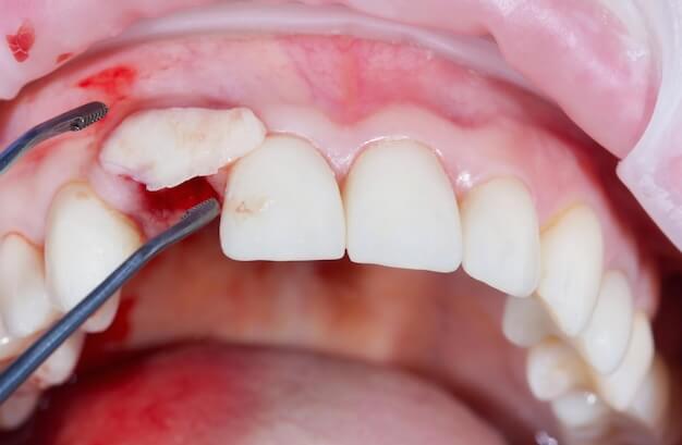
Gingival graft surgery is a periodontal surgical procedure performed to increase the thickness of the soft tissue surrounding the tooth and the amount of keratinized gingiva in areas where gingival recession and/or insufficient gingival tissue exist. The goal of this surgery is not simply to “lift the gum up.” From a broader perspective, the aims include the protection of the root surface, reduction of sensitivity, lowering the risk of bleeding and pain during brushing, making the gingival margin more stable, and decreasing the progression rate of recession over the long term. In some patients, aesthetic concerns are also an important reason; especially asymmetry in gingival levels in the anterior region can significantly affect smile aesthetics.
Gingival recession may be associated with many factors such as improper brushing technique (excessive pressure and hard-bristled brush), thin biotype (thin gingival structure), teeth positioned close to the alveolar bone boundaries, orthodontic movements, bruxism or teeth grinding, periodontal disease, smoking, and oral piercings or habitual trauma. Before planning graft surgery, these causes must be evaluated; because if the underlying overload or trauma continues, recession may recur after grafting. Therefore, graft surgery is often part of a “treatment plan” that includes oral hygiene education, periodontal cleaning, correction of traumatic brushing habits, and if necessary, occlusal or orthodontic adjustments.
Graft material is most often taken from the patient’s own tissue (autograft). The most common donor sites are the palate in the upper jaw (palatal area) or, in some techniques, the maxillary tuberosity region. Among the graft types used are free gingival graft, subepithelial connective tissue graft, and in some cases, alternative materials such as allografts or collagen matrices. The choice of technique depends on the class and depth of recession, gingival biotype, keratinized tissue requirements, root surface position, and the patient’s aesthetic expectations. In most cases, the objective is to maximize root coverage while also increasing tissue thickness and stability.
During the healing period, the care of both the grafted area and the tissue donor site is crucial. Sensitivity, swelling, and slight discomfort in the palate can be expected in the first days. The timing of suture removal varies depending on the suture type used but is generally planned between 7 and 14 days. Complete maturation of the soft tissue is a longer process; therefore, follow-up appointments, photographic measurements, and hygiene monitoring are integral parts of the treatment.
What Is Gingival Graft Surgery?
Gingival graft surgery is a soft tissue grafting procedure performed to increase insufficient gingival tissue around a tooth or to cover a root surface exposed by recession. The graft is mostly a piece of tissue taken from the patient’s own mouth, which is transferred to the target area and stabilized with sutures. Successful integration of the graft is closely related to the vascularization of the recipient site and control of micro-movement. Thus, postoperative care and avoidance of trauma play a critical role as much as the surgical technique itself.
This surgery has two main objectives: functional/protective and aesthetic. Functionally, exposed root surfaces can lead to increased sensitivity to cold and heat, easier root wear, and discomfort during brushing. Additionally, when keratinized gingiva is insufficient, the gingival margin becomes more sensitive and recession can progress. The graft aims to increase tissue thickness to make this area more resilient. Aesthetically, especially in the anterior region, the goals can include raising the gingival margin and improving symmetry.
The choice of graft type varies according to the goal. Free gingival grafts are preferred particularly to increase the band of keratinized tissue; although the tissue color may not perfectly match the surrounding gingiva, it is effective in achieving stable tissue gain. Connective tissue grafts, root coverage aim
Gingival Graft Surgery Procedure
The process begins with a detailed periodontal assessment. The depth of gingival recession is measured in millimeters, the classification of the recession is evaluated, the width of the attached/keratinized gingiva is measured, and the gingival biotype (thin/thick) is analyzed. If there is abrasion (cervical abrasion), decay, or restoration on the root surface, the plan is adjusted accordingly. Additionally, brushing technique, signs of tooth grinding, orthodontic history, and the tooth’s position within the alveolar bone are assessed. In some cases, photographs and measurement records are taken for more objective pre- and post-treatment follow-up.
The procedure is usually performed under local anesthesia. First, tissue preparation is done in the recipient site (the area with recession). Depending on the technique used, a tunnel can be created under the gingiva (tunnel technique), or a flap can be raised to expose the root surface. The root surface is cleaned of biofilm and irregularities; some protocols may use special agents for root surface preparation. Then, the graft donor site (usually the palate) is anesthetized locally, and the planned graft type is harvested. In connective tissue grafting, the palatal surface epithelium is preserved while the underlying connective tissue is taken; in a free graft, epithelium and connective tissue are harvested together.
The graft is transferred to the recipient area and secured with sutures. Since stabilization is critical, sutures are planned to prevent graft movement. In some cases, the graft surface is covered with a flap; this can support graft vascularization and aesthetic harmony. The donor site (palate) is also sutured to control bleeding or supported with a protective periodontal dressing or plaque. Written and verbal care instructions are provided to the patient at the end of the operation.
During the postoperative period, brushing in the recipient area is usually suspended for a certain time; instead, antiseptic mouth rinses and gentle hygiene methods are applied as recommended by the clinician. Nutrition is planned with soft foods, avoiding hot and irritating items. A control appointment is scheduled within the first week to evaluate tissue color, edema, and suture stability. Suture removal is generally planned between 7–14 days, depending on the suture type used and the healing rate.
Who Is Suitable for Gingival Graft Surgery?
Suitability for gingival graft surgery is evaluated based on the type of recession and the targeted gains. Patients with pronounced sensitivity to hot and cold, exposed root surfaces, pain/bleeding during brushing, and thin gingival tissue or insufficient keratinized gingiva band may be appropriate candidates. The lower anterior region and the upper canine–first premolar area are common sites of recession. Graft surgery can also be planned for patients with aesthetic concerns who want to improve gingival levels in the anterior region.
It is important that periodontal disease is under control. Performing graft surgery during active gingival inflammation, high plaque levels, and uncontrolled periodontal pockets may reduce success. Therefore, periodontal cleaning and hygiene education are provided first to stabilize tissue health. In patients with brushing trauma, improvement of brushing technique is necessary before surgery; otherwise, the new graft tissue may also be subjected to trauma.
The tooth’s position and interdental supportive tissues affect root coverage success. In certain recession types (especially those with significant interdental bone loss) complete root coverage may not always be possible; the goal can be stability and sensitivity control.
This surgery is a part of periodontal treatment. The plan includes not only covering the recession but also eliminating the factors causing the recession. With the right patient, correct technique, and good maintenance, long-term stable results are targeted.
Gingival Graft Surgery Preoperative Considerations
Preoperative preparation increases the likelihood of graft acceptance. The first step is to optimize oral hygiene. If there is inflammation or bleeding tendency in the surgical area, periodontal cleaning is performed first, and brushing and interdental cleaning techniques are corrected. Especially since hard-bristled brushes and horizontal brushing pressure may increase recession, a soft-bristled brush and correct technique are recommended. If signs of teeth grinding or clenching are present, management of this behavior, which increases load on the teeth and restorations, is planned; night guards may be recommended for some patients.
A detailed medical history should be evaluated. Blood thinners, diabetes medications, allergies, and medications affecting the immune system must be reported. Coordination with relevant physicians is provided if necessary. For procedures performed under local anesthesia, prolonged fasting is generally not required on the day of surgery; however, if sedation is planned, fasting protocols and accompaniment are necessary. Since sensitivity may persist for a few days after the procedure, planning work/school schedules is helpful.
Because the donor site will be the palate, the patient should be informed about a period requiring avoidance of hard and sharp foods that could increase palatal trauma. If the patient has stomach sensitivity or drug intolerance, the pain management plan should be adjusted accordingly. If the patient smokes, reducing smoking before surgery and abstaining afterward is an important step for graft success.
Expectation management is also necessary. Hundred percent root coverage may not be possible in every case; the success criteria are defined based on the intended functional gain (reduction of sensitivity, increase in tissue thickness, stabilization of recession) and aesthetic goals. Preoperative photos and measurements can help communicate these goals more clearly.
Gingival Graft Surgery Postoperative Considerations
The first few days after surgery are critical for maintaining graft stability and healthy vascularization. Mechanical trauma to the graft site (brushing, hard foods, touching with fingers) can displace the graft and reduce success. Therefore, the clinician may advise suspending brushing in the recipient area for a certain period and maintaining hygiene with antiseptic rinses. The rinse should be used as recommended in duration and frequency; early or forceful rinsing must be avoided. Mild soreness and sensitivity can be expected in the donor palate; nutrition should be planned to avoid irritating this area.
Bleeding, especially in the palate, may occur as oozing in the first hours. Contact the clinic if continuous fresh bleeding or inability to control the clot occurs. Pain medications prescribed should be taken regularly for pain management. Swelling is usually limited; however, cold application during the first 48 hours can increase comfort in some cases. Smoking negatively affects graft vascularization; abstaining during the healing period is critically important.
Nutrition should be adjusted to protect the graft: lukewarm and soft foods are preferred, and very hot, highly spicy, and acidic foods are avoided. Hard-shelled foods, chips, toast, and other sharp/firm foods may traumatize the palate and graft area. Ample fluid intake is important. If sutures are present, do not manipulate them. Suture removal is typically planned between 7 and 14 days postoperatively; this timing may vary depending on the case.
a) Achieving complete root coverage may not be realistic; in such cases, the goal can be set as increasing tissue thickness and reducing sensitivity. In patients undergoing or planning orthodontic treatment, the timing of the graft is planned by evaluating the effect of tooth movement on recession.
Systemic factors also influence suitability. Uncontrolled diabetes, smoking, immunosuppression, and certain medications can negatively affect wound healing. Smoking can reduce microcirculation and impede graft healing; therefore, smoking cessation or reduction is requested before and after surgery. An ideal candidate is someone who can comply with home care protocols, attend follow-up appointments regularly, and protect the area from trauma during the healing period. The Healing Process Depends on the Material Used and the Healing Process
During follow-up appointments, tissue color, graft adaptation, swelling, and signs of infection are evaluated. If any unpleasant odor, increased pain, fever, rapidly increasing swelling, or noticeable opening occurs in the graft area, a check-up should be done without delay. The maturation of the graft tissue continues over weeks; during this period, brushing technique is retrained and atraumatic hygiene is maintained. In the long term, regular periodontal check-ups and professional cleanings are important to keep the graft stable.
In Which Cases is Gingival Graft Surgery Performed?
Gingival graft surgery is performed for specific clinical indications. The most common indication is the exposure of the root surface due to gingival recession. This condition can cause sensitivity, aesthetic problems, and a risk of decay or abrasion on the root surface. When the band of keratinized gingiva is insufficient, the gingival margin becomes more sensitive and recession may progress; the graft can be applied to increase this tissue. Particularly in patients who experience pain while brushing near the cervical area of the tooth and struggle to maintain hygiene, comfort during brushing can improve after grafting.
Before or during orthodontic treatment, if the planned movement of the tooth is close to the bone border and the gingival biotype is thin, grafting may be planned to reduce the risk of recession. When keratinized mucosa around implants is insufficient, soft tissue grafts can be applied to facilitate implant care and increase soft tissue stability. In prosthetic planning, graft surgery may be considered in regions where soft tissue stability is important.
If tension occurs at the gingival margin due to frenulum pulling inside the mouth, frenectomy can be planned either together with the graft or in a separate session. Performing grafting without controlling factors such as traumatic brushing, tooth grinding, and periodontal disease may reduce long-term success; therefore, indication assessment is done alongside management of the causes.
Why is Gingival Graft Surgery Performed?
The graft surgery is performed to thicken the tissue, stabilize recession, reduce sensitivity, protect the root surface, and when necessary, to provide aesthetic improvement. In patients with a thin biotype, the gingival margin is more fragile and recession may progress even with minor trauma. The graft can make the gingival tissue more resilient and slow this progression. When the root surface is exposed, dentinal tubules come into contact with the external environment causing increased cold-hot sensitivity; root coverage or increased tissue thickness can reduce this sensitivity.
The exposed root surface is more prone to decay and can wear down during brushing. Gingival graft surgery aims to make the root surface more protected and reduce these risks. Moreover, as the patient’s comfort during brushing increases, long-term hygiene becomes more sustainable and this supports periodontal health. From an aesthetic perspective, especially in the anterior region, harmonizing the gingival levels can significantly improve the smile esthetics.
The long-term success of the graft depends not only on the surgery itself but also on managing the factors causing the recession. If traumatic brushing is not corrected or tooth grinding continues, the graft tissue may also be compromised. Therefore, graft surgery is often considered a “protective investment”: results are aimed to remain stable with correct habits and regular check-ups.
How Long Does Gingival Graft Surgery Take?
The duration of the surgery varies depending on how many teeth will receive grafts, the extent of recession, the chosen technique (connective tissue graft, free gingival graft, tunnel technique, etc.), the size of the graft harvested from the donor site, and any additional procedures performed in the same session. There is a difference in time between grafts applied to a single tooth area and those applied to multiple teeth. Management of the donor site (palate bleeding control, application of protective stent) and suture stabilization at the recipient site are important components of the surgical time.
In addition to the procedure time, the healing schedule must also be planned. The first week is critical for tissue stability. Follow-up check-ups are generally scheduled in the first week.
The removal of stitches can generally be planned between 7 and 14 days in most cases. The maturation of the soft tissue continues for weeks; during this period, brushing techniques are gradually reintroduced and follow-ups are maintained with professional cleanings. Complete aesthetic seating and tissue adaptation may take longer depending on the individual. The dentist clarifies both the post-procedure day plan and the follow-up schedule after the examination.
Gingival Graft Surgery Prices
Prices vary depending on the graft technique to be applied, the number of areas to be grafted, donor site management, the sutures and surgical consumables used, necessary periodontal preparatory procedures, the number of follow-up appointments, and additional requirements (such as frenectomy, root surface restoration). Soft tissue grafts around implants or extensive area grafting cases may increase the scope. In some cases, alternative graft materials (such as collagen matrix) may be considered, which can affect the treatment plan.
To obtain up-to-date and personalized information about gingival graft surgery prices, you should contact us. After the examination and periodontal measurements, once a personalized treatment plan is created, the cost information will be shared transparently.