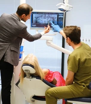

Soreness after dental extraction is normal, but a perforated sinus is an entirely different story that deserves proper attention. This article guides you through everything you need to know about sinus perforation following a tooth extraction, from identifying the earliest symptoms to understanding complications, prevention, and your path to recovery. If you’ve recently had an upper back tooth removed — or plan to soon — knowing this material could make all the difference between a smooth healing process and something far more troublesome. Let’s walk through every aspect and question with the kind of clarity and detail you deserve.
What Is a Perforated Sinus After Tooth Extraction?
When you extract a tooth in the upper jaw, especially those in the back (like the maxillary molars), the roots sometimes come quite close to the maxillary sinus cavity. The maxillary sinus is a hollow air-filled space located behind your cheekbones, lined with a delicate membrane. If the extraction process breaches this slim membrane, an opening may form between your mouth and the sinus — known as an oroantral communication, or more simply, a perforated sinus.
This isn’t just a theoretical risk: anatomical studies reveal that up to 13% of upper molar extractions result in some communication with the sinus, though most are minor and heal without surgical intervention. When the sinus membrane is punctured, bacteria, fluids, or air can pass between your mouth and sinus. The result can be a persistent open pathway that risks ongoing infection or chronic sinus problems. Even a “pinpoint” exposure can allow germs access to an area previously sealed off from oral bacteria.
Proper management is absolutely essential because unrecognized or poorly handled perforations can spiral into longstanding sinusitis, mouth-to-sinus fistulas, or even more complex infections demanding surgery. It’s rare, but the risks are real enough that every dentist is trained to spot and address this complication early on.
Causes of Sinus Perforation During Tooth Extraction
The roots of upper back teeth can extend close to, or even protrude into, the maxillary sinus. When an extraction is performed, several factors come into play that might cause a sinus perforation:
- Root Anatomy: Molars with long or divergent roots (often the first and second upper molars) are most likely to communicate with the sinus floor. In about 40% of the population, the roots of these teeth are separated from the sinus by only 1-2 millimeters of bone — barely thicker than a fingernail.
- Infection/Abscess: Chronic dental infections can erode the bone between the tooth and the sinus, dramatically thinning this barrier and making membrane perforation more likely during extraction.
- Surgical Technique: Excessive force or use of instruments like elevators in tight spaces can tear the delicate sinus lining. Failure to identify fused roots, root curvatures, or patient-specific risk factors increases the odds of a complication.
- Aging and Bone Loss: With age, bone around teeth naturally thins, and the sinus may expand lower into the jaw, especially when teeth have been missing for years. Osteoporosis and periodontal disease also reduce bone density.
- Anatomical Variation: Every patient’s sinus and root configuration is unique, so what works in one mouth might create a problem in another.
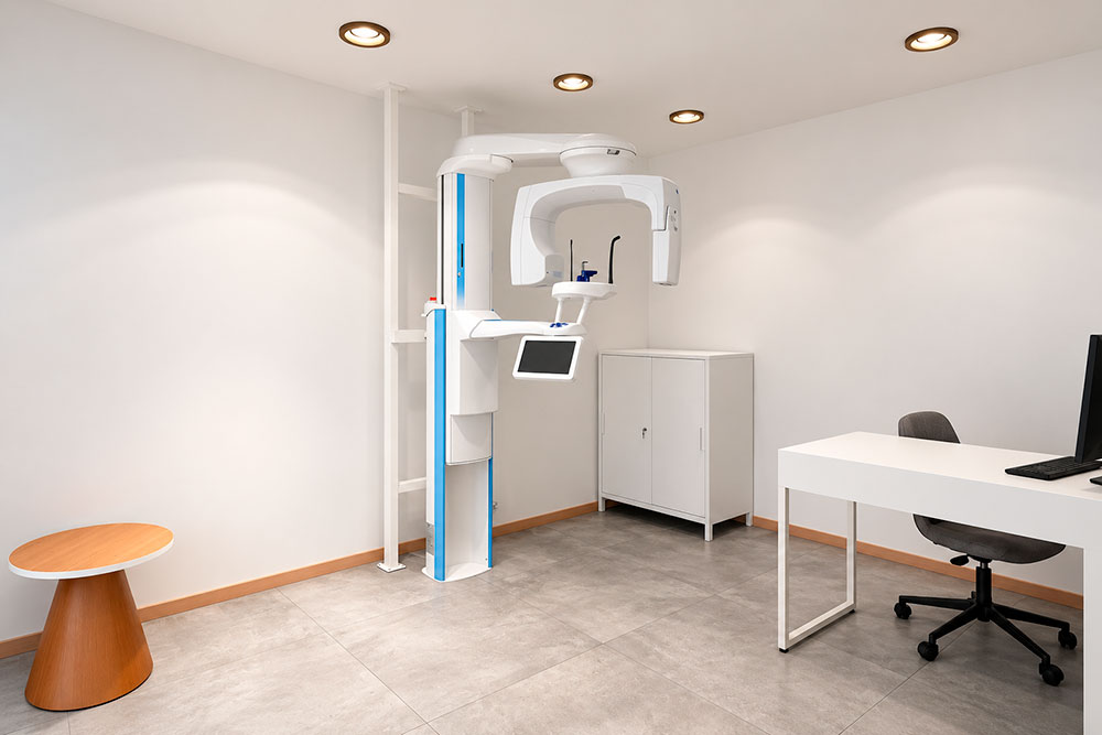
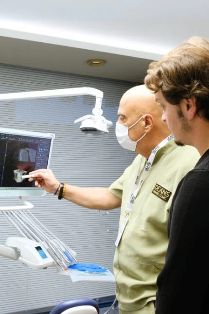
Pre-operative X-rays (usually panoramic, sometimes CBCT) help specialists assess risk before any extraction is performed. If high risk is noted, extraction technique may be modified, or additional repair materials may be kept ready in the operatory.
Early Symptoms of a Perforated Sinus
Spotting a sinus perforation early can save you a lot of discomfort down the line. Most individuals won’t realize what’s happening until certain symptoms make themselves known. Here’s what to watch for if you’ve had an upper molar or premolar extracted:
- Sudden or persistent passage of air/fluid: When you drink, you might notice liquid leaking through your nose or a “bubbling” sensation at the extraction site. Sometimes, simply breathing out can force air between your mouth and nose through the hole.
- Strange nose or cheek sensations: Watch for unexpected whistling, a feeling of fullness, or tingling in the cheek.
- Nasal voice quality: Your speech may suddenly sound more “nasal” than usual, especially when you’re eating or drinking.
- Unexpected bleeding: Gentle blows to the nose may prompt fresh bleeding at the extraction site, or blood might be visible when you spit after brushing teeth.
- Sinus pressure or headache: Early pressure or a dull ache in the cheek/upper jaw often signals something isn’t healing as it should.
If you notice any of these symptoms, especially leakage of fluid through your nose or the taste of sinus secretions at the surgery site, it’s time to contact your dentist right away. The sooner we step in, the higher the chance of simple, nonsurgical repair.
Complications Associated With Sinus Perforation
An untreated or poorly managed sinus perforation opens the door to complications that can dramatically affect quality of life. The most common is chronic oroantral fistula. That’s a persistent channel between your mouth and sinus, creating a direct pipeline for bacteria and debris to move in and out — never a good thing. Persistent oroantral fistulas can also make eating and drinking uncomfortable and embarrassing, as fluids and food particles may escape into the nose or sinus. Chronic sinusitis often develops in these cases, causing facial pain, congestion, post-nasal drip, and sometimes even repeated upper respiratory infections.
Another risk: oral antral communication can cause materials like food and dental debris to enter the sinus, resulting in a “foreign body” reaction that may require surgical removal. Over time, bone and soft tissue around the opening can become inflamed or infected, leading to foul taste, persistent bad breath, or even abscess formation that threatens neighboring teeth or bone.
Rare, but serious cases can see infection spreading through the sinus to the orbit or deeper facial structures, requiring hospitalization and intravenous antibiotics. The burden isn’t just physical; chronic sinus communication is emotionally taxing, too, as patients worry about social acceptance and are frequently forced to alter their diets and social activities.
Diagnosis of a Perforated Sinus
Experienced dentists rely both on what you report and on their clinical examination to make the diagnosis. Sometimes the perforation is apparent immediately — visible as an open hole at the base of the socket, or evidenced by air bubbling through the site when you breathe out with your nose pinched. In more subtle cases, a “Valsalva test” can help: you’re asked to close your mouth, pinch your nostrils, and gently blow. If air escapes through the extraction socket, that’s a clear indicator.
We typically use panoramic radiographs to check the relationship between the extraction site and the maxillary sinus. If there’s any doubt or if the perforation is suspected to be extensive, a cone-beam CT scan (CBCT) provides 3D imaging. These scans allow us to see both the size of the communication and whether any foreign objects (such as root tips or bone fragments) remain in the sinus itself.
Measurement of the perforation matters. Communication less than 2 millimeters typically heals spontaneously; anything larger, or complicated by infection, needs intervention. Diagnostic accuracy here is non-negotiable — missing a sinus perforation is one of the surest ways to guarantee future complications. We take great care not to rush these assessments.
Treatment Options for Sinus Perforation
Treatment depends on both the size of the perforation and when it’s caught. Small holes (<2mm) often require nothing more than careful instruction to avoid nose-blowing, sneezing with the mouth closed, and creating any negative pressure for 2–3 weeks. We usually prescribe saline rinses and sometimes a short course of antibiotics as a preventative measure. For moderate defects (2–6mm), a collagen plug or resorbable membrane is often placed in the socket. Overlying stitches pull the gum tissue closed, effectively sealing the hole. Soft food and oral hygiene instructions follow.
Larger communications, or those present for longer than a week without closure, may require more advanced flap surgery. A common method is the buccal advancement flap: we mobilize tissue from the cheek side of the socket, advance it over the perforation, and suture it in place. This provides immediate coverage and a high likelihood of full closure. Some surgeons use a palatal rotation flap or combine membranes with biologically active bone grafting agents if bone is missing.
If infection is present within the sinus (evidence by persistent pain, foul discharge, fever), targeted antibiotics are started, and sometimes referral to an ENT for sinus drainage is necessary. Chronic cases may require closure by oral-maxillofacial surgeons under sedation or general anesthesia. Most repairs (even surgical ones) are completed in under 45–60 minutes and performed right in the dental operatory. Long-term follow-up is mandatory until complete healing is confirmed, usually within one to two months.
Prevention Strategies During Tooth Extraction
Preventing sinus perforation begins before the extraction even takes place. High-resolution radiographs (like CBCT) help map out root proximity and recognize thin sinus membranes. For at-risk teeth, we sometimes perform “sectional” tooth removal — breaking the tooth into pieces and extracting roots with minimal force rather than one large pull, reducing the risk of membrane tear.
Meticulous use of elevators and forceps, with the gentlest technique possible, is standard. In some cases, I’ll create a tiny window in the bone to remove roots atraumatically — a method known as a “surgical extraction.” Maintaining good patient positioning and keeping the area well irrigated also protects the sinus lining. If the sinus is inadvertently entered during extractions, immediate closure using a resorbable membrane or soft tissue flap is performed while you’re still numb, drastically reducing later complications.
Finally, communicating risk to patients — so you know what to monitor — closes the loop. When both doctor and patient act proactively, the odds of major sinus problems fall dramatically.
When to See a Dentist or Oral Surgeon
Contact your dentist or oral surgeon if you’ve had a recent upper tooth extraction and notice:
- Persistent leakage of fluid from the mouth to the nose or vice versa
- Unusual nasal or cheek swelling after the first 48 hours
- Whistling sounds, air passage, or bubbling at the extraction site
- Persistent bleeding, particularly with nose blowing
- Sinus pain, facial pressure, or ongoing bad taste/breath
Do not attempt to self-treat with over-the-counter sinus or allergy products. Prompt intervention within days of the perforation can often spare you from more involved, surgical repairs. If symptoms continue or worsen, seek in-person evaluation. Where in doubt, it’s always safer to get checked early.
Frequently Asked Questions (FAQ)
What are the most common symptoms of a perforated sinus after tooth extraction?
The classic symptoms include feeling or hearing air movement through the extraction site when you breathe out, sudden nasal leakage of fluids when drinking, or a bubbling sensation at the socket. Patients frequently report a strange nasal voice, unexplained bleeding, or a sense of fullness in the cheek. Some individuals feel pain or pressure that persists beyond the typical healing timeline. If you ever blow your nose and blood seeps from the extraction area, this is a hallmark sign. Taken together, these symptoms should prompt immediate contact with your dental provider for evaluation.
How long does it take for a perforated sinus to heal after dental extraction?
Healing timelines depend on the size and management of the perforation. Small perforations under 2mm often close spontaneously within two to three weeks, provided no negative pressure (like nose blowing) is generated. With protective closure (sutures and membrane placement), moderate-sized openings heal within 2–4 weeks for most patients. For surgically repaired, larger connections, complete tissue integration can take four to six weeks, sometimes longer if bone grafting is required. We usually schedule follow-up visits every 10–14 days to monitor healing, repeating imaging or performing a “Valsalva” test as needed until the connection is fully resolved.
Can a perforated sinus heal on its own without treatment?
Very small communications (less than 2mm) often do heal without surgical procedures, especially if identified early and the patient avoids forceful nose blowing, sneezing with the mouth shut, or sucking through straws. We always caution patients to heed post-extraction instructions precisely in these cases. Larger communications, those complicated by infection, or any fistula persisting more than a week almost always require active closure by a dental specialist. Don’t gamble with hope — if symptoms persist, intervention is generally needed, and waiting too long increases the complexity of what’s required for repair.
What risks are associated with untreated sinus perforations?
Leaving a sinus perforation untreated can lead directly to chronic oroantral fistula — a permanent passage between mouth and sinus. This increases the risk for ongoing sinusitis, recurrent infections, and social embarrassment (leakage of fluids, food debris entering the sinus, or foul odors). Over time, inflammation can impact bone and adjacent teeth, degrade oral health, and, in rare instances, allow infection to reach deeper facial structures that are much more difficult to treat. Quality of life often declines as chronic discomfort, repeated doctor visits, and self-consciousness become persistent issues for affected patients.
How is a perforated sinus diagnosed by dentists?
Dentists use a combination of clinical history, physical exams, and imaging to confirm the diagnosis. Commonly, a Valsalva maneuver is performed — the patient pinches their nose and gently blows to see if air passes through to the mouth. Visual inspection may reveal an open passage at the site of extraction. Panoramic X-rays or, for more detailed mapping, cone-beam CT (CBCT) scans help us see the size and precise location of the communication. The process is straightforward and non-painful, with accuracy greatly improved by documenting symptoms promptly and clearly during your visit.
What are the most effective treatments for a perforated sinus after tooth extraction?
Small perforations close spontaneously with conservative care: gentle oral hygiene, no nose blowing, and possibly a prescribed antibiotic. When a larger opening is discovered, we frequently place a collagen plug or resorbable membrane, and close the gums tightly with sutures — fast, effective, and minimally invasive. More substantial or persistent communications may require a soft tissue flap such as a buccal advancement flap. Sinus infections complicating the picture require antibiotics; in rare cases, ENT referral for sinus lavage or surgical drainage may be needed. The majority of cases are resolved successfully with timely dental management, usually in under a month from repair.
Is sinus perforation during tooth extraction common?
While not an everyday occurrence, sinus perforation is a recognized risk – especially with upper molars and premolars. Scientific studies report incidence rates ranging from 0.5% to 13% for these teeth, with wide variation depending on patient age, anatomy, and the presence of chronic infection. Most perforations are very small and heal on their own, but a smaller subset requires targeted treatment. Dentists are trained to minimize risk via careful radiographic and clinical assessment pre-operatively, so with modern best practices, serious complications are becoming less common than in decades past.
What should I avoid if I suspect I have a perforated sinus after extraction?
Avoid anything that could force air or fluids through the extraction site. This means no nose blowing, stifling sneezes (always sneeze with your mouth open), no forceful sucking through straws, and holding off on smoking or vaping, which can disrupt the fragile healing tissue. Try not to spit vigorously or drink carbonated beverages in the first few days. Heavy lifting or bending over for long periods should also be minimized, as increased sinus pressure can delay closure. Follow every instruction given by your dentist and return for all recommended follow-up exams to ensure healing is progressing on track.
Can sinus infection develop from a perforated sinus after tooth extraction?
Yes, a sinus infection (sinusitis) is a leading complication when oral bacteria gain access to the maxillary sinus via the open communication. You might experience persistent facial pain/pressure, thick nasal discharge (often with a foul odor), headache, and sometimes fever. Infections can become chronic if not treated promptly, spreading to adjacent spaces and requiring more aggressive salvage therapy. This is why both antibiotic prophylaxis and timely closure of the perforation are considered standard care by dental professionals when a sinus breach is detected.
Are there surgical procedures to repair severe sinus perforations?
Yes, severe or persistent sinus perforations are most effectively treated with surgical repair. The buccal advancement flap remains the gold standard; tissue is freed from the side of the jaw and advanced over the hole for watertight closure. Palatal or combined flaps and placement of resorbable membranes may be chosen for larger defects or where initial repairs have failed. Bone grafting may be included if significant bone loss is detected. Intractable or infected cases sometimes require joint oral-maxillofacial and ear/nose/throat (ENT) specialist intervention with sinus debridement and closure under sedation. Success rates for surgical closure exceed 95% with current best practices, and most procedures are completed as outpatient treatments with minimal downtime for the patient.